Aside from a 12-lead ECG placement theres something known as a 15-lead placement which includes placing leads V4-V6 on the posterior side of the patient below their left scapula see below. Lead Placement for Posterior ECG Resus Review.
Lead Placement For Posterior Ecg Resus Review
It is also helpful for future clinicians if you note in your read that it is a posterior ECG.
. Position trainer in the desired upright or horizontal position. 2 patients among the 50 had both RVI and PWMI. Aside from a 12-lead ECG placement theres something known as a 15-lead placement which includes placing leads V4-V6 on the posterior side of the patient below their left scapulasee below.
Isolated posterior MI is less common 3-11 of infarcts. Posterior infarction accompanies 15-20 of STEMIs usually occurring in the context of an inferior or lateral infarction. Posterior ECG leads.
Basic 12-Lead Placement 1. Traditional 15 Electrode Placement Two Channel 5 Electrode Lead Placement In this conguration two channels of ECG data are bipolar. Continuing Medical Education 12 Lead ECG interpretation Practice 12 lead 3 You are called to an office building where a 45 year old executive is complaining of chest pain after a very stressful situation.
While the 18-lead ECG is perhaps more sensitive for early detection of ischemia or infarction in practice either should be used for. A posterior wall MI even though the initial 12 lead ECG shows no obvious acute changes The fact that it doesnt directly show up on a standard 12 lead ECG is the reason the posterior wall MI is the most. Ensure the trainer is clean.
Hints of an associated posterior infarct. The standard 12-lead ECG does not assess these areas directly Consequently. Besides the incidence of isolated posterior MI is not defined and has been reported in studies ranging from 0 to 7-12 18 23.
To detect posterior infarcts which are often associated with inferior or lateral wall AMI. They are performed by placing V4 V5 and V6 electrodes in the same intercostal space but continuing into the patients back. 15 or 18 lead ECGs can be done with alternate precordial lead placement to assess for posterior- or right-sided disease.
Amongst these 10 patients had posterior MI 56 con-firmed on 15 leads ECG. Posterior extension of an inferior or lateral infarct implies a much larger area of myocardial damage with an increased risk of left ventricular dysfunction and death. Lead ECG taken from 50 IWMI patient s the overall incidence of ST elevation in the posterior chest.
Doing a 15 lead ECG. See figures 8 9 3. RS amplitude ratio in V1 or V2 is 1.
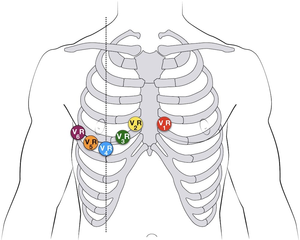
A complete set of right-sided leads is obtained by placing leads V1-6 in a mirror-image position on the right side of the chest see diagram below. Leads V7-V9 was 26. Suspected right ventricular or posterior infarcts.
ST depression in V1 and V2 with R waves. In addition the use of the 15-lead ECG confirms the posterior MI and is superior to the findings in the anterior leads. Isolated posterior MI is less common 3-11.
Red positive is referenced to white negative and brown positive is referenced to black negative Poster AHA IEC Label Color Color Channel Lead Location. In this series of 15 -. When viewing the EKG strip V4-V6 on the strip will be referred to as V-13-15.
STD in V1-V3 or. Firstly do a standard ECG then by repositioning leads V4 V5 and V6 to the patients back they become V7 V8 and V9. V4 - V7 should be placed at the level of V6 at the posterior axillary line.
12-Lead ECG Interpretation Introduction This self-study package has been developed to provide a review of twelve lead interpretation as well as a review of signs and symptoms of various types of AMIs. Posterior infarction accompanies 15-20 of STEMIs usually occurring in the context of an inferior or lateral infarction. Terior leads ECG were also taken.
2-3 Level A Recommendation When a 15-lead or 18-lead ECG machine is not available manipulation of the leads from a standard 12-lead ECG machine allow additional areas of the heart to be imaged4-5 Indications of a posterior wall infarction may include4-513 Changes in V 1 V 3. Lead Placement for Posterior ECG. ST-elevation myocardial infarction STEMI is suspected when a patient presents with persistent ST-segment elevation in 2 or more anatomically contiguous ECG leads in the context of a consistent clinical history.
Presenting with suspected Posterior Myocardial Infarction PMI To determine the utility of 15-lead ECG in the early diagnosis of acute posterior myocardial infarction Backgroun d 7 Acute posterior myocardial infarctions PMI and right ventricular myocardial infarctions are likely to be underdiagnosed. 15 lead ECG was the diagnostic modality in 100 of patients in this study. There are three situations where a 15 lead ECG should be performed after a 12 lead ECG.
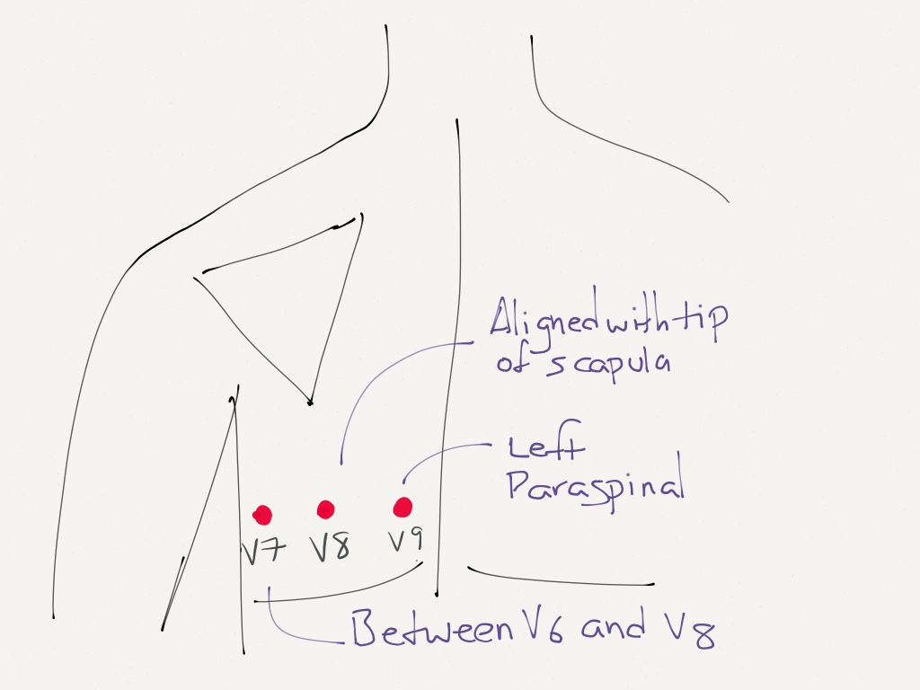
V5 -V8 at the tip of the scapula. Lay out labeled leads and plug them into their designated outlets on the 15-lead electronics box. Where do you place a 15 lead ECG.
The last time I did a posterior EKG was on a guy who told me he last had a posterior wall MI. Proper 12-Lead ECG Placement. Electrodes Placement for Posterior Leads.
On most EKg machines the labels areno automatically changed so it is important to cross out the labels for V4-V6 and write in V7-V9. Ill do a right 15 or 18 lead if Im really suspicious of something cardiac going on but cant immediately find it on a 12 lead or if I see an inferior wall MI. 12- 15- lead ECG Section 1.
In the fifth intercostal space and the left posterior axillary line. When viewing the EKG strip V4-V6 on the strip will be referred to as V-13-15. V4V7 V5V8 and V6V9.
Posterior ECG leads V7-V9 are applied by moving V4-V6 to under the left scapula. To clarify leads will equal. The leads V4-V6 are removed and substituted for V7-V9 as shown below.
Continuing Medical Education Section 1. You suspect that the underlying cause of a patients presentation is cardiac eg. Accuracy of 12 leads ECG for detection of posterior.
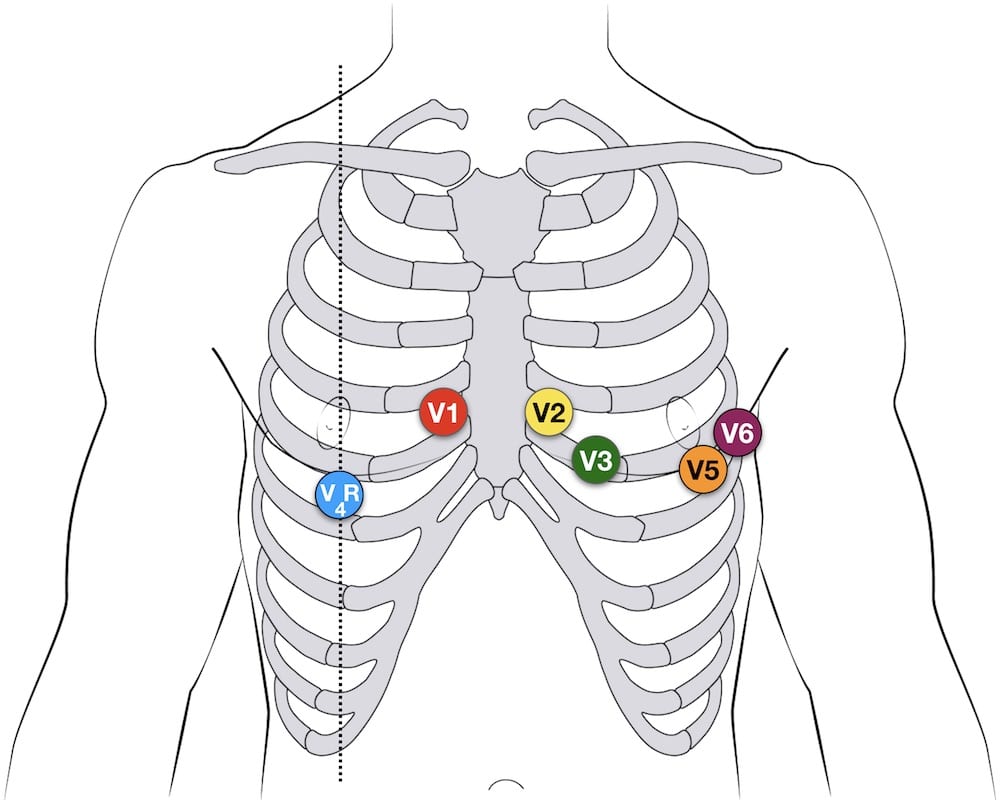
Posterior leads are helpful in suspected posterior myocardial infarction. Feel for anatomical landmarks on trainer remove electrode from sheet and place adhesive side. It can be simpler to leave V1 and V2 in their usual positions and just transfer leads V3-6 to the right side of the chest ie.
Right sided 12 lead ECG lead placement. Out of total of 176 patients 18 patients 1022 had changes suggestive of posterior MI on 12 leads ECG. Aside from a 12-lead ECG placement theres something known as a 15-lead placement which includes placing leads V4-V6 on the posterior side of the patient below their left scapula see below.
Ecg Lead Positioning Litfl Ecg Library Basics

How To Not Miss A Posterior Myocardial Infarction Em Daily

Diagnostics Alternative Ekg Leads Taming The Sru

Aliem Cards

Ecg Educator Blog Posterior Ecg Lead Placement
Emdocs Net Emergency Medicine Educationposterior Mi Recognition Emdocs Net Emergency Medicine Education

Ecg Lead Positioning Litfl Ecg Library Basics
Ecg Lead Positioning Litfl Ecg Library Basics
0 komentar
Posting Komentar